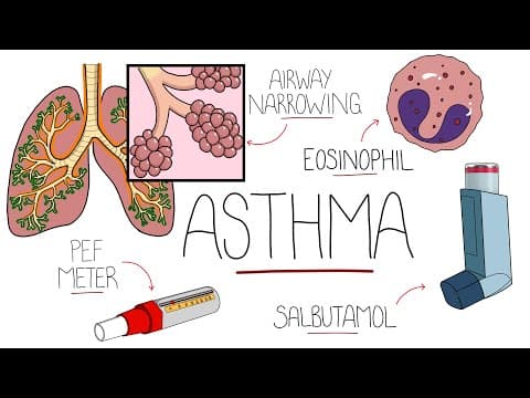
[0:01]Asthma, which comes from the Greek word meaning panting, is a chronic inflammatory disease affecting the lower airway, characterized by hyperresponsiveness and bronchospasm, leading to a reversible narrowing of the airways that can be life-threatening. The conduction zone of the airways, which extends from the nose to the bronchioles, is where air is warmed and moistened before getting to the respiratory zone, where gases are exchanged at the alveoli. In asthma, it is the conducting zone that is mostly affected, in particular, the bronchi and bronchioles. Looking at a bronchus more closely, there are multiple layers including the lumen where air is present, a mucus layer to help protect against foreign particles, the epithelial cells, lamina propria, and smooth muscle cells. Now, we'll see why these are especially important in the pathophysiology of asthma. The first phase of an asthma exacerbation is the early phase, happening in the first few minutes. In asthma, there is thought to be an excess of T-helper 2 cells. This is also true in allergic dermatitis, known as eczema, and allergic rhinitis, known as hay fever, which is why these three together make up the atopic triad. Allergic asthma is the most common type of asthma, triggered by allergy to things like pollen, house dust or mites, and pet antigens. But there are also non-allergic triggers such as smoking and even perfumes. The trigger is taken up by an antigen presenting cell, typically a dendritic cell, and shown to the T helper cell. In asthmatic patients, these TH2 cells produce cytokines in response, including IL4, IL5 and IL13. IL4 and IL13 cause plasma cells to release IgE, and IgE will then activate mast cells to release granules in a process known as degranulation. These granules include histamine, leukotriene, and prostaglandin. In immunology, this is called a type one hypersensitivity reaction. The release of these molecules leads to the contraction of the smooth muscle layer in the airways, known as bronchospasm, an increased production of mucus and also edema, all together giving a narrower airway, which produces the asthma symptoms. IL5 leads to activation of eosinophils, which also release more cytokines and leukotriene, which contribute to this process as well. The release of all these inflammatory mediators leads to more inflammatory cells being recruited from the blood over the next few hours, known as chemotaxis, and further production of inflammatory mediators, giving the second phase of the exacerbation, termed the late response. These changes are initially reversible, but as these exacerbations happen over years, the airway becomes remodeled and is no longer entirely reversible. The changes include sub-epithelial fibrosis and basement membrane thickening, hypertrophy of the smooth muscles, larger volume of mucus coming from goblet cell hyperplasia and also increased vascularity. Asthma is thought to affect 235 million people worldwide, with around 1 in 12 adults in the United States being affected. Multiple factors come together to cause asthma. For example, genetically, it is thought that having a parent with asthma makes you between three and six times more likely to develop it. Environmental factors can affect not only exacerbations but also the development of asthma. The hygiene hypothesis proposes that having reduced exposure to pathogens at a young age can increase the TH2 response and predispose to asthma. Linked to this is the association with cesarean section delivery and a 20 to 80% increase in the risk of asthma. Other factors include smoking during pregnancy and living with low air quality, and a history of atopic disease is the strongest risk factor. The characteristic findings in asthma are recurrent episodes of shortness of breath or dyspnea, wheezing, chest tightness, and coughing. These are often worse at night or in the early morning and can also be worse in response to cold air and exercise. Anxiety is also more common in patients with asthma, as are obstructive sleep apnea and gastroesophageal reflux disease, which is often misdiagnosed as asthma. We mentioned that the airway may become permanently remodeled, and this can lead to chronic obstructive pulmonary disease, known as COPD, a disease that is frequently seen in smokers. There is also an increased prevalence of anxiety and depression in people who suffer from asthma. Often overlooked, it's important to remember that asthma exacerbations can lead to respiratory failure and death. The initial diagnosis of asthma is made on the clinical history and symptoms, and may be confirmed using spirometry, where the volume of air inhaled and exhaled is measured. The main values looked at are the forced expiratory volume in one second and the forced vital capacity. Overall, the patient takes a maximum breath in and then blows out as hard and fast as possible and continues until no more air is expired. The FEV shows the volume of air expired in one second, while the FVC shows how much air can be forcefully exhaled in one breath. The ratio between these shows the proportion of air that a person is able to expire in one second compared to their forced vital capacity. This is normally around 0.75 or 75%. In obstructive respiratory conditions, like asthma and COPD, we expect that this ratio is lower than 0.75. If the FEV1 improves by over 12% after receiving bronchodilators, this is supportive of a diagnosis of asthma due to reversibility. Peak expiratory flow also measures the volume of air exhaled in a quick exhalation and can be measured at home using a peak expiratory flow meter.
[7:05]Asthmatic patients are encouraged to do the peak expiratory flow when healthy, and then the expiratory flow achieved during an exacerbation can be compared. To diagnose acute exacerbations, it is mostly down to the clinical symptoms, but there are features that are suggestive of a more severe exacerbation, which can be remembered with a pneumonic, A-chest. A is for an altered conscious level or arrhythmias, C for cyanosis and CO2, which if normal or high is worrying in asthma, as it may be a sign of fatigue. H is for hypotension and hypoxia, E for exhaustion from fighting to breathe, and S is for a silent chest due to severe broncoconstriction, meaning not enough air flow is present to generate wheezing. T is for a threatening peak flow, which is defined as below 33% of their best. While there is no definitive cure for asthma, there are treatments to help reduce symptoms and exacerbations, which can also help to reduce airway remodeling. Long-term management of asthma largely involves a cycle of assessing the current situation, such as identifying risk factors, adjusting them or introducing medication, and then reviewing the response. This often means a step-wise strategy for medication. A salbutamol inhaler may be used as a reliever and may be the only therapy necessary. However, if symptoms persist, then a low dose inhaled corticosteroid is introduced. From here, the next step is mostly introducing a long-acting beta2 agonist like salmeterol and increasing the dose of inhaled corticosteroid. Leukotriene receptor antagonists like montelukast can also be introduced. At the highest level, there may be the addition of long-acting muscarinic antagonists like tiotropium, and the use of targeted therapy, like anti-IgE such as omalizumab, or anti-IL4 or IL5 therapies. For acute exacerbations, oxygen is usually given, and relieving therapy like short-acting beta2 agonists and anti-cholinergic like ipratropium are used by nebulizers. Systemic steroids are also used, which is typically prednisolone. If these measures do not resolve the exacerbation, magnesium sulfate intravenously is another option. But in severe cases, mechanical ventilation may be required. Viral infections are more commonly the trigger for acute exacerbations. However, if a bacterial cause is suspected, then antibiotics are started.


