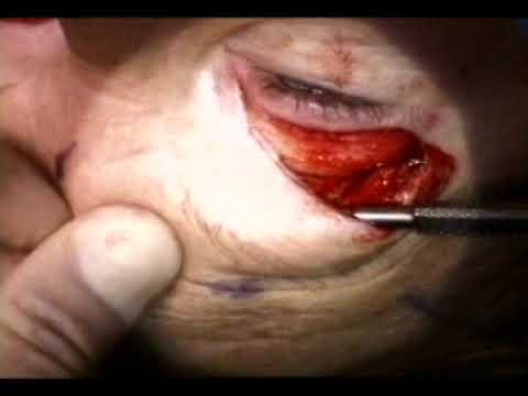
[0:00]This is Richard Allen at the University of Iowa. This video demonstrates an endoscopic brow lift using a camera on the surgeon's headlight for visualization. The view during this video will be from the surgeon's view and upside down compared to other videos shown. An incision is made with the monopolar cautery along the blepharoplasty markings. A flap of skin and orbicularis muscle is removed. The medial fat pad is mobilized. The fat pad is then prolapsed forward and conservatively excited.
[0:37]Dissection is then carried out superiorly between the orbicularis muscle and the orbital septum to the superior orbital rim. The superior orbital rim is then identified and the periosteum is incised with the needle tip cautery. The periosteum is then elevated from the underlying bone with a freer periosteal elevator.
[1:06]Medially, the super orbital neurovascular bundle is identified and preserved. Attention is then directed to the scalp incisions. A 15 blade is used to make an incision through the scalp to the underlying bone. A freer periosteal elevator is then used to elevate the periosteum from the underlying bone. The periosteum is then elevated inferiorly. Laterally, it is elevated to the conjoined tendon. This is performed with the gold handled elevator. Usually the elevation is performed blindly to the roof of the nose. I believe that this is safe. Attention is then redirected to the blepharoplasty incisions where additional periosteal elevation is performed medially. This is performed with the super orbital muscular bundle in mind and protected. Using a freer periosteal elevator, one can then demonstrate that the forehead is completely mobile. Attention is then directed to the other side where the periosteal elevation is performed in the exact same manner.
[2:27]Attention is then directed to the temporal incisions. These incisions straddle the conjoined tendon. An incision is made with the 15 blade through the skin and subcutaneous fat. The superficial temporalis fascia is then identified and incised. The deep temporalis fascia is then identified and metzenbomb scissors are used to dissect along the surface of the deep temporalis fascia. A freer periosteal elevator is then used to find the subperiosteal pocket medial to the conjoined tendon. Metzenbaum scissors are then used to lyse the tendon between the sub periosteal pocket medially and the pocket along the surface of the deep temporalis fascia laterally. Attention is then redirected to the scalp incisions where the endotine drill is used to make a hole into the bone. The endotine forehead device is placed through the incisions and popped into the bone.
[3:38]This is then released and the scalp is elevated over the endotime. Prior to doing this, the freer periosteal elevator is used to ensure that the endotine is not caught on scalp before elevation. After elevation, the surgeon's thumb is used to push down on the endotime.
[4:02]The same procedure is then performed on the opposite side.
[4:09]Attention is then directed to the temporal incisions where the superficial temporalis fascia is engaged with a 3-0 vicril suture. This suture then engages the superficial temporalis fascia superiorly.
[4:25]Tying the suture results in plication of the superficial temporalis fascia and therefore a smash lift. The same procedure is performed on the other side. Again, engaging the superficial temporalis fascia with 3-0 vicryl suture, then the needle engages superficial temporalis fascia superiorly. I believe that just one suture is necessary in this area. I do not engage deep temporalis fascia, but many surgeons will.
[5:01]The scalp incisions are then closed with staples. I believe staples give an excellent cosmetic closure and sutures are not needed here.
[5:13]The eye incisions are then closed with a running 6-0 prolene suture. At the conclusion of the case, the patient is cleaned with wet and dry gauze, erythromycin and ophthalmic ointment is placed over the eye incisions. Bacitracin ointment is placed over the scalp incisions, a headband is placed for two days and the patient returns in approximately one week.


