[0:35]Hello everyone. Uh today I will talk about the small intestine and the pancreas. Okay. Uh
[0:57]Okay, again. So, we'll talk about the small intestine and the pancreas. Start by the small intestine. Okay. Small intestine is about 6 meters. It extends from here, from the pylorus, of the stomach. And it terminates here, the ileocecal junction. So, it extends from the pylorus of the stomach till the ileocecal junction. Small intestine is divided into three parts: duodenum, the first 10 inches, 25 centimeters. Then the Jejunum, proximal two-fifths of the small intestine. And then the Ilium, the distal or the lower three-fifths of the small intestine. Actually the Jejunum is located above the umbilicus, and the Ilium is located below the umbilicus. Okay. So, uh, we have to compare between the Jejunum and Ilium. The Jejunum is the proximal two-fifths of the small intestine. The Ilium is the distal three-fifths of the small intestine. The Jejunum is located above the umbilicus. The Ilium is located below the umbilicus. The Jejunum is reddish in color because it is more vascular. The wall is more vascular. The Ilium is pale in color because it is less vascular than the Jejunum. The wall of the Jejunum is thick due to the presence of these mucous folds. Several, many, numerous mucous folds called plicae circularis. Look at the wall of the Ilium. The wall of the Ilium, uh, looks smooth, okay, or thin, due to absence of mucous folds, or the presence of few mucous folds. Actually, the distal part of the Ilium, uh, contains, doesn't contain, uh, doesn't contain mucous folds. Okay? Again. The Jejunum arteries, these are the Jejunum arteries, are simple, or form simple arcades, form one to two arcades. The Jejunum arteries, the Ileal arteries are complicated, and form three to four complicated arcades. These are the arcades, okay? And regarding the mesenteric fat, this is the fat. The, uh, uh, the mesenteric fat of the Jejunum part, is a small amount forming windows. You can see the arcades. The mesenteric fat in the Ileal part of the mesentery, contains large amount of fat, so forming no windows. So, Jejunum arteries are simple. Ileal arteries form complicated arcades, three to four arcades. Mesenteric fat is less in the Jejunum part. And mesenteric fat is large in the Ileal part. The Jejunum, regarding lymphoid follicles, lymphoid follicles are absent in the upper part of the Jejunum and are few in the lower part of the Jejunum. But in the Ilium, there are numerous, uh, lymphoid follicles, and they may aggregate to form Peyer's patches. Okay.
[4:54]Then we'll talk about the mesentery of the small intestine. This is the mesentery of the small intestine, which suspends the small intestine from the posterior abdominal wall. It is fan-shaped. Okay? It is fan-shaped because the free border is long, and the root or the attached border is short. Okay? The mesentery of the small intestine, it extends from the duodenojejunal flexure till the ileocecal junction. It has two borders, free border, which surrounds small intestine, and the attached border, and the attached border, which is attached to the posterior abdominal wall, and it's called the root of the mesentery. Then we'll talk about the relations and the contents of the mesentery. Okay, this is the mesentery.
[6:03]It crosses the fourth and third part of the Jejunum. Then it crosses three main structures, large structures.
[6:16]This is the abdominal aorta, then the inferior vena cava, then the right psoas muscle and the ureter. So, regarding the relations of the mesentery, or the root of the mesentery. The root of the mesentery crosses four structures: the duodenum, the abdominal aorta, the inferior vena cava, the right psoas with the right ureter. Then we'll talk about the contents of the mesentery. This is the mesentery. Okay? This is the free margin or the free border, and this is the attached border. So, the contents. Number one, Jejunum and Ileum in free border.
[7:03]Jejunum in the upper part, Ileum in the lower part. Okay? Jejunum and Ileum in the free margin. This one, superior mesenteric vessels in the root. Between the root and the free margin, you can see these branches from the superior mesenteric artery, which form arcades, simple arcades in the upper part, complicated arcades in the lower part. So, Jejunum and Ileal branches forming arcades. From the terminal branches, vasa recta supply the intestine.
[7:56]In the mesentery, sympathetic fibers around the blood vessels. Mesenteric lymph nodes, arranged in three layers, small size at the free margin, medium size in the middle, and large size at the root. Then extraperitoneal fat or mesenteric fat. Mesenteric fat, less in the Jejunum part forming windows. And dense in the Ileal part, no windows. So, again, contents of the mesentery: Jejunum and Ileum in free border. Superior mesenteric vessels in the root. Jejunum and Ileal branches forming arcades. From the terminal branches, vasa recta supply the intestine.
[8:58]Sympathetic fibers around the blood vessels. Okay, this is the mesentery, small intestine in the free margin. This is the Jejunum, this is the Ileum. Superior mesenteric vessels in the root. Jejunum and Ileal branches, okay, forming arcades. Simple arcades in the Jejunum part, complicated arcades in the Ileal part. Lymph nodes, small size, medium size, large size. Mesenteric fat is less in the Jejunum part, dense in the Ileal part. Okay? Remember, this is the Ileum, this is the Jejunum, this is the Ileum. How do you identify the Jejunum from the Ilium? By the presence of these mucous folds, plicae circularis.
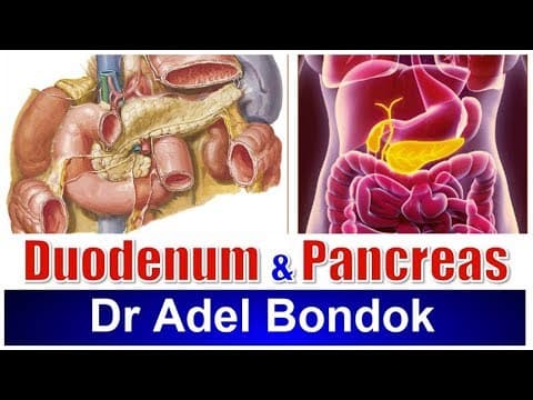
[10:23]Then we'll talk about the duodenum. Okay? Regarding, it is 10 inches long. It is divided into four parts: first part, second part, third part, and fourth part. Remember 2-3-4-1, or 1-2-3-4. I add one inch to the number of the part, so the first inch, the first part is two inches, the second part is three inches, the third part is four inches, but the fourth part is only one inch.
[11:12]The first part lies in the transpyloric plane opposite L1. The third part lies in the subcostal plane opposite L3. The second part, which is the vertical part, extends from L1 to L3. And the fourth part extends from L3 to L2. Then we'll talk about the relations, then we'll talk about the relations of the first part. This is the first part of the duodenum. This is the first part of the duodenum. So, it is two inches long. Then we'll talk about the relations. It is opposite L1, the transpyloric plane. It is partially covered with peritoneum, okay? The first inch is mobile because it has lesser omentum and greater omentum. The first inch, like the stomach, the second inch is partially covered with peritoneum. Okay? So, the peritoneal covering of the first part. Or actually, the peritoneal covering of the whole duodenum. The whole duodenum is partially covered with peritoneum except the first inch. Which is completely covered with peritoneum like the stomach, because it has lesser omentum and greater omentum. So the mobile part of the duodenum is the first inch. Then we'll talk about the relations of the first part. What is anterior and what is posterior? Okay? What is above and what is below? So, anterior relation, this is the first part of the duodenum. Anterior relation, we have two structures, these two structures. Quadrate lobe of the liver and gallbladder. Okay? Then posterior relation. This is the hepatic artery. Okay? Posterior relation. Remember, in the free margin of the lesser omentum, we have the bile duct, common bile duct. This is the bile duct. Okay? Traveling behind the first part of the duodenum, then behind the head of the pancreas. The hepatic artery gives a branch behind the first part of the duodenum, gastroduodenal artery. Very good, which divides into right gastroepiploic and superior pancreaticoduodenal. Remember, behind the bile duct and the hepatic artery is the portal vein. And behind the portal vein is the inferior vena cava.
[14:10]Therefore, behind the first part of the duodenum, bile duct, gastroduodenal artery, portal vein, inferior vena cava. But actually, because the first inch is related to the lesser omentum and greater omentum, so behind the first inch is the lesser sac. Then what is above? Above the first part of the duodenum is the epiploic foramen. And below the first part of the duodenum is the head of the pancreas. Again, relations of the first part of the duodenum: in front of the first part of the duodenum, gallbladder and quadrate lobe of the liver. Behind the first part of the duodenum, this is the bile duct. Gastroduodenal artery, portal vein, inferior vena cava. Superiorly, it is the epiploic foramen, inferiorly it is the head of the pancreas. Then the second part of the duodenum. Again, just reminding you of the structures related to the first part of the duodenum, behind the first part of the duodenum. This is the hepatic artery, okay? This is the bile duct. Okay? So, behind the first part of the duodenum, this artery, gastroduodenal artery. Behind the artery and the duct, portal vein, behind the portal vein, inferior vena cava. Okay. Now the second part of the duodenum. This is the second part of the duodenum. It is three inches long. As I said before, the whole duodenum is partially covered with peritoneum except the first inch. So, partially covered with peritoneum except the area crossed by the transverse colon, which is bare. Okay? Relations. The second part extends from L1 to L3. Yes. Then what is anterior to the first, to the second part? Three structures. The upper part is related to the liver. Right lobe of the liver. The middle part is crossed by the transverse colon. Here, transverse colon. And the lower part is related to the small intestine. Okay? So, the second part of the duodenum is related anteriorly to three structures. Liver, right lobe of the liver, transverse colon, okay, and small intestine. Behind the second part of the duodenum is the hilum of the right kidney, and the right psoas muscle. Okay? Medially, it is the head of the pancreas. And the head of the pancreas is separated from the second part of the duodenum by two arteries: superior pancreaticoduodenal and inferior pancreaticoduodenal arteries. And laterally, it is the right colic flexure. And you should know the structures which open in the second part of the duodenum. Structures which open in the second part of the duodenum. We have two papillae: major duodenal papilla and minor duodenal papilla. The major duodenal papilla is formed by the common bile duct and the main pancreatic duct. And the minor duodenal papilla is formed by the accessory pancreatic duct. Okay? So, structures opening in the second part. This is the major duodenal papilla. And this is the minor duodenal papilla. The major duodenal papilla in the middle of the second part, is formed by opening of bile duct and the main pancreatic duct. The accessory pancreatic duct opens in the minor duodenal papilla, one inch above the major.
[18:32]Then the third part of the duodenum, it is four inches long. Again, it is partially covered with peritoneum. Okay? Third part of the duodenum, it lies opposite the subcostal plane, opposite the third lumbar vertebra. What is anterior and what is posterior? Anterior, we have three structures.
[19:00]This root of the mesentery.
[19:08]And remember, the artery which crosses the third part of the duodenum, superior mesenteric vessels. And loops of small intestine. Okay? Posterior, three main structures separated by another three structures.
[19:31]So, the three main structures: aorta, separated by the inferior mesenteric artery. Then inferior vena cava, separated by the right gonadal artery. Then the right psoas muscle, separated by the right ureter. So, the three main structures: aorta, inferior vena cava, right psoas. The three other structures: inferior mesenteric artery between the duodenum and the aorta, right gonadal artery between the duodenum and inferior vena cava, right ureter between the duodenum and the psoas muscle. Still have superior, head of the pancreas.
[20:24]Then the fourth part of the duodenum, which is one inch long and is partially covered with peritoneum.
[20:34]The duodenojejunal flexure.
[20:44]Is attached to the right crus of the diaphragm, or connected to the right crus of the diaphragm, by the suspensory ligament of Treitz. This one.
[20:58]Then we'll talk about the arterial supply of the duodenum. I mentioned earlier that the duodenum develops from the foregut and the midgut. Therefore, it is supplied by two arteries. The part above the biliary orifice is supplied by the celiac artery because it is developed from the foregut. The part below the biliary orifice is supplied by the superior mesenteric artery because it is developed from the midgut. Therefore, the duodenum is supplied by the upper half by the celiac artery. And the lower half is supplied by the superior mesenteric artery. What are the branches which supply the duodenum? Okay? From the celiac artery, right gastric, this one, right gastroepiploic.
[21:59]From the gastroduodenal, and this one, supraduodenal, and this one, superior pancreaticoduodenal. So actually, if you look at these four arteries, actually these four arteries are branches from the hepatic artery. So the upper part of the duodenum is supplied by the hepatic branch of the celiac artery. What are the branches of the hepatic artery which supply the upper part of the duodenum? Right gastric, right gastroepiploic, supraduodenal, superior pancreaticoduodenal. The lower half of the duodenum from the superior mesenteric artery, from this branch, inferior pancreaticoduodenal artery. Nerve supply and lymph drainage. Same is applied for the nerve supply and lymph drainage. This is the area crossed by the transverse colon bare area, okay? So, nerve supply. The pancreas is supplied by the celiac plexus except the lower part of the head and uncinate process. So, nerve supply. The upper half of the duodenum is supplied by the celiac plexus. This is the celiac plexus. The lower half is supplied by the superior mesenteric plexus. Lymph drainage, same. The upper half drains into the celiac lymph nodes. The lower half drains into the superior mesenteric lymph nodes.
[24:06]Then the pancreas. Okay? Position of the pancreas. It extends from the concavity of the duodenum to the hilum of the spleen, in the left hypochondrium.
[24:30]It is divided into four parts: head, neck, body, and tail.
[24:38]Then we'll talk about the relations of the pancreas. Arterial supply, nerve supply, lymph drainage, and the pancreatic ducts. Main pancreatic duct and accessory pancreatic duct. Okay, let us start by the head of the pancreas.
[25:01]Position of the head. The head of the pancreas is located in the concavity of the duodenum. It has this hook-like process, it's called uncinate process. So, the head of the pancreas is located in the concavity of the duodenum. It has uncinate process. Then we'll talk about the relations of the head of the pancreas. What is anterior, what is posterior? What is superior, what is inferior? This is very simple. Okay? Then anterior. Anterior to the head of the pancreas, remember the second part of the duodenum is crossed by the transverse colon. Transverse colon, superior mesenteric vessels in front of the uncinate process.
[26:10]What is posterior to the head of the pancreas? Remember the bile duct, inferior vena cava, aorta actually behind the uncinate process and behind the neck.
[26:43]Neck and uncinate process.
[26:48]Okay? Laterally, talking about the head of the pancreas, it is the second part of the duodenum. Separated from the head of the pancreas by superior and inferior pancreaticoduodenal vessels. Superiorly, the first part of the duodenum. Inferiorly, it is the third part of the duodenum. So, the head of the pancreas, crossed anteriorly by the transverse colon, the uncinate process is crossed anteriorly by superior mesenteric vessels. Behind the head, bile duct, inferior vena cava, aorta actually behind the uncinate process. Okay? Laterally, second part of the duodenum, separated from the head of the pancreas by superior and inferior pancreaticoduodenal vessels. Then the neck of the pancreas. This is the neck of the pancreas. Actually, this is the neck of the pancreas. The neck of the pancreas is the landmark for two structures. Origin of two structures.
[28:04]Origin of the portal vein. Origin of this artery, superior mesenteric artery.
[28:18]Okay? So, this is the neck of the pancreas. So, relations of the neck of the pancreas. What is anterior to the neck of the pancreas? Lesser sac and first inch of duodenum. Gastroduodenal artery.
[28:40]Posteriorly, three veins and two arteries.
[28:47]Talking about the neck of the pancreas.
[28:52]What are the three veins?
[28:56]Of course this one, splenic vein.
[29:01]Yes, termination of the splenic vein.
[29:08]And this vein, termination of superior mesenteric vein, and beginning of the portal vein. Two arteries. Of course this one, behind the neck of the pancreas.
[29:26]Aorta, and origin of superior mesenteric artery. So, behind the neck of the pancreas, three veins: termination of the splenic vein, termination of superior mesenteric vein, beginning of the portal vein. Two arteries: aorta and origin of superior mesenteric artery. Then the body of the pancreas. This is the body of the pancreas. This is the neck. Okay?
[30:00]Actually, the body of the pancreas is triangular in cross section. This is upper border, this is lower border, this is anterior border. The upper border is related to the splenic artery. The anterior border gives attachment to the transverse mesocolon. This is the anterior surface, this is the inferior surface, and this is the posterior surface. Okay? The anterior surface.
[30:50]The inferior surface is related to the greater sac and small intestine, Jejunum. Then the posterior surface crosses these structures.
[31:17]Left crus of diaphragm, sympathetic chain, right psoas, sorry, left psoas, left suprarenal gland, left kidney.
[32:20]Three veins: splenic vein, inferior mesenteric vein, and left renal vein.
[33:31]Then the tail of the pancreas. This is the tail of the pancreas.
[33:40]The tail of the pancreas, it passes in this ligament, lienorenal ligament. Yes.
[33:51]It is related to the visceral surface of the spleen below the lateral end of the hilum.
[34:03]Arterial supply of the pancreas. The pancreas is supplied by the celiac artery except the lower part of the head and uncinate process. So, celiac artery except the lower half of the head and the uncinate process. By superior mesenteric artery. So, the head of the pancreas, upper part of the head, supplied by superior pancreaticoduodenal artery. Lower part of the head and uncinate process, by the inferior pancreaticoduodenal artery. Neck, body, and tail by the splenic artery.
[34:47]Nerve supply and lymph drainage. Think about the blood supply. So, nerve supply. The pancreas is supplied by the celiac plexus except the lower part of the head and uncinate process. So, celiac plexus, except the lower half of the head and the uncinate process, which are supplied by superior mesenteric plexus. Lymph drainage, same. Upper half of the head, neck, body and tail to the celiac lymph nodes. Lower half of the head and uncinate process to the superior mesenteric lymph nodes.


