[0:03]I'm going to begin with an overview of hemostasis first. I'm trying an endothelial cell, and the purpose of hemostasis is to stop any bleeding right away when there's injury to the endothelial cell.
[0:16]The way we do this is first we make a platelet plug. And this is the goal of primary hemostasis.
[0:22]And then, in order to make that stronger, what we do is we link fibrin together on top of that platelet plug and make a mesh.
[0:31]And this is what makes the platelet plug stronger.
[0:37]And this is what we accomplished, this is what we do in secondary hemostasis.
[0:44]And when we have primary and secondary working together and we form this mesh with the platelet plug, this is called a clot.
[0:49]Right now, I'm just going to focus on primary hemostasis.
[0:53]I'm going to put the endothelium cell up to the right and to the side, so that we remember that we're focusing on the platelet plug.
[0:59]I'm going to bring in another endothelium cell that we're, we'll work with.
[1:04]And I'm going to cause some damage, cause some injury and see what, what, um, what our bodies do in order to stop that bleeding.
[1:11]The first step in primary hemostasis is vasoconstriction. What we want to do is clamp down.
[1:17]The smooth muscle cells in the blood vessel want to clamp down and make the hole that blood blood is flowing through smaller.
[1:27]So I'm drawing it right now, and you can see that, that the, the hole that the blood can flow through is smaller.
[1:33]And this is going to decrease the amount of, of blood that we lose.
[1:38]The way I like to think about it is, say we're on a bridge and we're driving and all of a sudden half the bridge on one side collapses.
[1:45]In order to stop any cars from falling off or limiting the amount of damage, the police come right away and they slow down traffic and they also probably cut down lanes.
[1:59]So they go from four to two in order make sure that the cars don't fall off.
[2:03]So that's what that's what the blood vessels are doing.
[2:08]We do the, we do vasoconstriction in two ways. One is just a nerve reflex. It's like a knee jerk reaction.
[2:16]We have some injury and then all of a sudden our nerves tell our smooth muscle cells to contract.
[2:19]A second way we do it is by this blue, blue molecule called endothelin.
[2:24]And it's secreted from the endothelial cells and acts on the smooth muscle cells in the blood vessel and causes vasoconstriction.
[2:33]It causes the smooth muscle cells to contract.
[2:36]But in order to know how endothelin does this, we need to talk about healthy endothelial cells and what healthy blood vessels do.
[2:44]So normally, on a healthy blood vessel, we're secreting all three molecules.
[2:50]The green molecules are nitric oxide and prostacyclin.
[2:56]And these are vasodilators.
[2:59]And endothelin, like I mentioned, is a vasoconstrictor.
[3:04]And they kind of play a tug of war with each other.
[3:07]Then the the cells are always secreting these substances, um, but vasodilation always tends to to win out in healthy cells, healthy blood vessels.
[3:17]And that makes sense because we want to make sure that blood is flowing through.
[3:20]But what happens during an injury to our blood vessels is that we have we lose the amount of nitric oxide and prostacyclin that we make.
[3:29]So endothelin wins over because there's no more nitric oxide in prostacyclin. So we're going to get vasoconstriction.
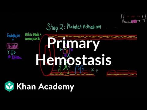
[3:38]And now, after vasoconstriction, we get platelet adhesion, meaning we need the platelets to to stick to the side of injury.
[3:46]We need the platelets together. In order to understand how the platelets do that, let's talk a little bit more about the platelets.
[3:53]I'm going to draw a platelet and normally they're not square and normally they're not that big.
[4:02]But I just want to make sure that we get a good picture of what's going on.
[4:05]So platelets are normally flowing around in our blood with red blood cells.
[4:09]And they carry around these two granules and they're called.
[4:14]Um, I like to think of them as sacks or sort of like backpacks on a camping trip.
[4:20]You carry around a lot of things in your backpack on that camping trip and you may not use any of it, but you carried it around just in case you might need it.
[4:28]That's how these granules work in these platelets. I want to talk about two receptors also.
[4:33]The platelets have more receptors, but there are two key ones that, that are important for primary hemostasis.
[4:40]The blue and the purple receptors that I'm drawing, they're both glycoproteins, but that's such a long name.
[4:48]So I'm just going to refer to them as their second part of their name.
[4:53]And I'm also going to write everything that we are talking about on the side, sort of like a scorecard.
[5:00]That we keep track of all the, all the molecules and receptors that are important for primary hemostasis.
[5:08]So the blue receptor is glycoprotein 1B, and the purple receptor is glycoprotein 2B3A.
[5:19]We'll just refer to them as 1B2B3A. Oh, sorry.
[5:25]Not B. It's 2B3A.
[5:28]Now, in order for platelets to find the side of injury, we have to talk about normal platelets first and their interaction with endothelium cells.
[5:35]Normally, platelets don't adhere or stick to endothelium cells.
[5:40]And this has to do with nitric oxide and prostacyclin.
[5:49]The two substances that we had already mentioned that are secreted by healthy endothelium cells.
[5:54]And what they do in addition to causing vasodilation to the smooth muscle cells is they kind of block the platelets from sticking to the endothelial cells.
[6:00]Like we mentioned, when there's injury to the endothelial, then there's less nitric oxide and less prostacyclin that will block platelets from getting closer to the endothelial cell.
[6:13]So now that there's less nitric oxide and prostacyclin, platelets are going to get closer to the, that side, the side of injury because there's nothing blocking it from going there.
[6:23]In addition to that, we also have this glue, this molecule that provides the link to the side of injury and to the platelet.
[6:33]This glue is called von Willebrand Factor.
[6:38]I wish there was a better way to remember that.
[6:41]It's a long name, and we'll just refer to it as VWF.
[6:45]And this VWF is normally floating around in our blood.
[6:50]But it also gets secreted from endothelium cells at the side of injury specifically.
[6:56]And when von Willebrand Factor comes into contact with the side of injury, it binds to subendothelial collagen.
[7:05]Collagen is a substance that provides structure to the blood vessels and it normally doesn't have contact to blood or to platelets.
[7:14]So at this injury, von Willebrand factor binds tightly to the collagen, and on the other side it binds to the GP1B receptor on the platelets.
[7:23]So the platelet is ready to bind von Willebrand factor when there's a side of injury.
[7:29]So now that it binds, once it's bound, then it actually the platelet actually gets activated and that's when we begin the next step, activation and degranulation.
[7:39]So when the platelet gets activated, it changes shape.
[7:44]So I'm erasing the platelet now, so that I can change the shape of it.
[7:51]And it also does many other things. One of them is the GP2B3A receptor is in a confirmation that is normally inactive.
[8:00]So it, it's not able to bind properly. And so after activation, it changes shape so that it's able to bind.
[8:08]And then the sacks, the granules that I mentioned that are inside the platelets, now they, they become of use and that's when degranulation happens.
[8:18]These sacks, these granules get released into, into the blood.
[8:22]One of them is called the alpha granule, and in the alpha granule, we have two substances that we already know about.
[8:30]One of them is fibrinogen, which we will be using in secondary hemostasis, and the other one is von Willebrand factor.
[8:39]And in the dense granule, which I like to think of it as dense and so it has more, and so it has this one has three substances, three molecules.
[8:52]And since we're thinking of them as backpacks or sacks, um, it also helps me remember what's in the dense granule.
[9:00]In the dense granule, in the dense sack, we have serotonin, ADP and calcium.
[9:07]And the way I remember what the three molecules do, I think of past, present and future.
[9:14]So, serotonin is, is released and it's a constructor of the smooth, smooth muscle cells.
[9:22]And so I think of that as past because that's what we did in the past, but now it's going to do it again.
[9:29]And ADP is present. ADP activates platelets and promotes aggregation.
[9:36]So ADP is what we need now in order to get that plate to clump, platelets to clump together.
[9:42]And then calcium is the future because calcium is needed for secondary hemostasis.
[9:47]That's the next part of making stabilizing that platelet plug.
[9:50]The third thing that an activated platelet does is secrete thromboxane A2.
[9:55]Thromboxane A2 is actually the op exact opposite of prostacyclin and it's made by the same enzyme.
[10:04]And thromboxane, plate also plays a tug of war with prostacyclin.
[10:08]It acts on smooth muscle cells to cause vasoconstriction and it also causes more platelets to activate and helps with aggregation.
[10:11]And so the final step, platelet aggregation is mediated primarily through GP, GP2B3A.
[10:20]And it's not until an activated platelet actually causes the 2B3A receptor to change to a shape that allows it to bind fibrinogen.
[10:30]Because 2B3A on the platelet binds fibrinogen, and it's through fibrinogen binding many 2B3A receptors from many platelets that creates the clumping and the platelet plug that we get at the end of primary hemostasis.


