[0:00]Welcome to the pain relief educational channel. Today we'll explore three essential techniques for performing cervical medial branch block using ultrasound guidance.
[0:11]Whether you are an interventional pain physician, anesthesiologist, or spine specialist, mastering these approach can significantly enhance accuracy and patients outcome. Let us start.
[0:24]This is a technical report exploring ultrasound techniques for cervical medial branch blocks offering a comprehensive analysis of three distinct needles approaches.
[0:35]Whether you are an experienced practitioner seeking to refine your technique or an inspiring professional integrating ultrasound in your practice, this presentation will provide invaluable insight to enhance your skills in interventional pain management.
[0:51]This presentation is based on an cadaveric study published in the pain physician journal in 2024.
[1:00]The lead author, Doctor Agnes Stogza collaborated with a team of experienced pain specialists to conduct this research.
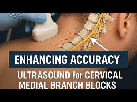
[1:08]Cervical medial branch blocks target the medial branch nerves that innervate the facet joints.
[1:16]These nerves are responsible for transmitting pain signals from the cervical spine, often due to facet joint arthritis or whiplash injuries.
[1:26]Blocking these nerves hold diagnostic and therapeutic significance. Ultrasound offers a radiation free alternative to fluoroscopy.
[1:38]With a real-time soft tissue visualization, it always used to identify critical landmarks like the articular pillars and medial branches without need for ionized radiation.
[1:48]The study objective is to assess and compare the precision and safety of the commonly used ultrasound guided cervical medial branch block techniques.
[2:00]The first approach is the coronal long axis technique where the authors place the probe in a longitudinal orientation to visualize the articular pillar.
[2:15]Medial branch located in the debi's point between the articular pillars.
[2:20]Of course, except for the third occipital nerve, which located at the C2-C3 joint. The needle is introduced from anterior to the ultrasound probe and slowly advance perpendicular to the beam.
[2:35]The needle is advanced from anterior to posterior because the vertebral artery and the neuro foramen are situated anterior to the facet line.
[2:45]The second approach is transverse short axis technique.
[2:49]The ultrasound probe is placed in a slightly oblique transverse plane to visualize the cervical articular pillar and the lamina.
[3:02]The needle introduced in a in plane technique from posterior to anterior until bony contact was made at the deepest part of the articular pillar for C3 to C6 and the C2 C3 joint for third occipital nerve.
[3:19]This method provides a direct needle path and may be preferred for certain anatomical variation. Third approach involved placing the needle as in the second approach.
[3:32]Then adjusting the needle tip using the ultrasound visualization of the C-spine in a coronal view.
[3:39]The author hypothesize that combining these two views might enhance needle precision. Following needle placement, fluoroscopic examination was performed using both anteroposterior and lateral views.
[3:55]Fluoroscopic image of each needle were then independently evaluated to assess placement accuracy and procedural safety.
[4:03]There are four key evaluation questions addressed in the study. First one, is the needle at the correct level, or has the level been missed?
[4:14]Determining whether the needle is appropriately positioned at the target cervical level. The second question is, is there is a correct crude placement?
[4:26]For the third occipital nerve, the needle should be within the joint line area. For C3 to C6 medial branches, the needle should be on the articular pillar within the designated black box.
[4:38]Third question is, is the needle precisely placed? The ideal needle tip position is within the target area, the green box here and here.
[4:51]Ensuring optimal therapeutic effectiveness. Fourth question, is the needle placed potentially dangerous?
[4:58]Evaluate whether the needle is in the compromising critical structure such as spinal cord content, existing nerve root, or vertebral artery. The collective results of the study are summarized in the following table.
[5:13]Target level were missed in 10.9%, 12%, and 10% of cases for the three approaches respectively.
[5:25]However, the difference between these methods were not statistically significant.
[5:31]No significant difference was found in the crude needle placement across the three approaches. Precise needle placement was significantly higher with the First approach compared to the other two approaches.
[5:44]The First approach also result in a statistically significant increase in the dangerous placement, raising the safety concern. The author stated that, giving the variability in the position of the medial branch, relative to the bony articular pillar,
[6:00]the reliability of the green zone as an ideal target area may be questioned. Additionally, the commonly used 0.3 ml injection volume for diagnostic block, typically diffused across the entire articular pillar at the given level, potentially reducing the importance of precise location.
[6:19]In conclusion, currently, the practitioner should be able to achieve a similarly successful block using the three CMBB described methods.
[6:30]However, the coronal long axis approach appears more beneficial in precisely positioning the needle at the centroid of the articular pillar.
[6:40]Despite this, it also increased the risk of placing the needle tip in potentially hazardous locations. I hope this walkthrough has provided valuable insight to help refine your techniques and enhance your confidence in performing cervical medial branch block under ultrasound guidance.
[6:59]Precision and safety are key in interventional pain management, and mastering this approach can make a real difference in patient outcome.
[7:07]If you find this video helpful, don't forget to give it thumbs up and share it with colleagues who might benefit and hit the subscribe button for more expert lead content on advancing pain procedure. Let us continue learning and improve together. Thanks for watching and I'll see you in the next video.


