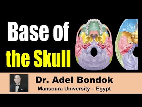
[0:15]Hello everyone. This is Dr. Adel Bondok, Professor of Anatomy and Neuroscience, Mansoura University, Egypt. I am going to discuss the base of the skull from outside and from inside, from the cranial cavity. I will start by Norma Basalis externa, okay. Normal base of external is divided into three parts. The anterior part is the hard palate. The middle part between the hard palate and anterior margin of the foramen magnum. And the posterior part behind the anterior margin of the foramen magnum. It is the occipital bone and the mastoid process. Start by the anterior part or the hard palate. So we'll talk about the formation of the hard palate, the sutures and the foramina. The hard palate is formed of two bones, the anterior two-thirds formed by the maxilla, palatine processes of the maxilla. The posterior one-third by the palatine bone, of course right and left. So, anterior two-thirds maxilla, posterior one-third palatine bone. And this spine or projection is the posterior nasal spine. Regarding the sutures and foramina, this suture in the midline is the median palatine suture. Okay? Median palatine suture. This suture between the maxilla and palatine bone is the palato-maxillary suture. This palato-maxillary suture is also called transverse palatine suture. We have three foramina, this is the first one in the midline, incisive fossa or incisive foramen. It transmits nasopalatine nerve and sphenopalatine vessels. This is the greater palatine foramen, transmitting the greater palatine nerve and vessels, and these foramina, small foramina, lesser palatine foramina, transmits lesser palatine nerve and vessels. So the hard palate, anterior two-thirds maxilla, posterior one-third palatine bone. Median palatine suture, transverse palatine suture. Incisive foramen, transmitting nasopalatine nerve and sphenopalatine artery. Greater palatine foramen, transmitting greater palatine nerve and vessels, lesser palatine foramina, transmitting lesser palatine nerve and vessels. Then the middle part of Norma Basalis. We'll talk about the bones in the midline and then on each side. In the midline here, this is the first one, part or the posterior part of the nasal septum, is called the vomer. So, this is the vomer, the posterior part of the nasal septum and then the sphenoid body of the sphenoid bone. It is not shown here. And this suture between the body of the sphenoid and the basilar part of the occipital bone, is called the occipitosphenoid suture. This occipitosphenoid suture ossifies at the age of 25 years. This is the basilar part of the occipital bone in front of the foramen magnum and this projection is the pharyngeal tubercle. So in the midline, the vomer, body of the sphenoid, occipitosphenoid suture, basilar part of the occipital bone, pharyngeal tubercle. On each side, this one and this one, this plate and this plate, medial and lateral pterygoid plates. Medial pterygoid plate and lateral pterygoid plate. Between the two plates is the pterygoid fossa. This fossa pterygoid fossa, this fossa pterygoid fossa. The medial pterygoid plate has two features. This is the pterygoid hamulus and this is the scaphoid fossa. Pterygoid hamulus, scaphoid fossa. Then we'll move laterally. This bone is the greater wing of the sphenoid. The greater wing of the sphenoid contains two foramina on the outer surface. This is foramen ovale and this is foramen spinosum. Foramen ovale transmits four structures that read male. Mandibular nerve, accessory meningeal art, lesser petrosal nerve, and emissary vein. Foramen spinosum transmits artery and nerve. The artery is the middle meningeal artery and the nerve is the nervus spinosus.
[6:02]Therefore, the middle meningeal artery passes through foramen spinosum. The accessory meningeal artery passes through foramen ovale. Okay? And then this groove is for the auditory tube, eustachian tube. This bone, this one, is the petrous part of the temporal bone. This petrous part of the temporal bone showing at the apex foramen lacerum. This foramen lacerum transmits internal carotid artery and deep petrosal nerve. The deep petrosal nerve arises from the sympathetic fibers around the internal carotid artery. This foramen is the carotid canal and the carotid canal extends between this foramen and foramen lacerum, inside the petrous part of the temporal bone. This foramen between the petrous part and the occipital bone, jugular foramen, this one. Okay? Move laterally. This is the styloid process. Okay? And this is the articular tubercle and articular fossa. The articular fossa is called mandibular fossa because it articulates with the head of the mandible to form the temporomandibular joint. Therefore, norma basalis externa, the middle part, in the midline, vomer, body of sphenoid, occipitosphenoid suture, basilar part of the occipital bone, pharyngeal tubercle. Medial pterygoid plate, lateral pterygoid plate, pterygoid fossa, okay? Pterygoid hamulus, scaphoid fossa. Okay? Greater wing of the sphenoid, foramen ovale, foramen spinosum. Petrous part of the temporal bone, foramen lacerum, carotid canal, jugular foramen, styloid process, articular tubercle and articular fossa. Then the posterior part of Norma Basalis externa is formed of occipital bone and mastoid process. This is the occipital bone and this is the mastoid process. Regarding the occipital bone, it is divided by the foramen magnum, this one, into three parts. So divided by foramen magnum into three parts. Basilar part anterior in front of the foramen magnum.
[8:48]Squamous part behind the foramen magnum and condylar part, two occipital condyles on each side of the foramen magnum. The squamous part shows this projection, external occipital protuberance. And then external occipital crest, superior nuchal line, and inferior nuchal line. Two foramina. One above the occipital condyle and one behind the occipital condyle. The one above is the hypoglossal canal. This hypoglossal canal is also called anterior condylar canal. It transmits the hypoglossal nerve.
[9:40]This is the posterior condylar canal. This posterior condylar canal transmits emissary vein between the sigmoid sinus and suboccipital venous plexus. Regarding the mastoid process, it shows this notch, mastoid notch. This mastoid notch is also called digastric notch because it gives attachment to the posterior belly of the digastric. This foramen is very important one, between the styloid and mastoid process. Stylomastoid foramen. This stylomastoid foramen transmits facial nerve and stylomastoid artery.
[10:37]And the stylomastoid artery is a branch from the posterior auricular artery and the stylomastoid artery supplies the facial nerve. So stylomastoid foramen is very important one. It transmits facial nerve and stylomastoid artery. This foramen is the mastoid foramen. This mastoid foramen is present at the occipitomastoid suture, mastoid foramen. It transmits artery and vein. The artery is the meningeal branch of the occipital artery and the vein is emissary vein between the sigmoid sinus and occipital veins. And this groove is made by the occipital artery. Okay, just a summary of Norma Basalis externa. This is the anterior part, hard palate. This is the middle part and this is the posterior. Start by the anterior part, which is the hard palate, formed of maxilla, forming the anterior two-thirds. Palatine bone forming the posterior one-third and this is the posterior nasal spine. This suture in the midline, median palatine suture, and this one is the palatomaxillary suture or transverse palatine suture. This is the incisive fossa or incisive foramen, transmits nasopalatine nerve and sphenopalatine artery. This is the greater palatine foramen and these are the lesser palatine foramina. Okay? This is the middle part. Start by here, this is the vomer. This is the basilar part of the occipital bone. This is the pharyngeal tubercle. This is the occipitosphenoid suture ossifies at the age of 25. Okay? Medial pterygoid plate and lateral pterygoid plate. Between the two, pterygoid fossa. The medial pterygoid plate related to two structures. Okay? Pterygoid hamulus and scaphoid fossa. And this is the groove for the auditory tube. Okay? The greater wing of the sphenoid shows two foramina, foramen ovale and foramen spinosum. This is the articular tubercle and articular fossa. This is foramen lacerum at the apex of the petrous temporal bone. This is the carotid canal and jugular foramen. This is the styloid process.
[13:59]This is the mastoid notch. This is the groove for the occipital artery. This is the mastoid foramen and this is the posterior condylar canal. And this is the anterior condylar canal or the hypoglossal canal.
[14:34]Then Norma Basalis interna or the cranial cavity. The cranial cavity is divided into three parts: anterior cranial fossa, middle cranial fossa, and posterior cranial fossa.
[15:06]Let us discuss each one. This is the anterior cranial fossa and this is the middle cranial fossa. We'll identify the bones and foramina. Anterior cranial fossa in the midline, this is the frontal crest, this is the foramen cecum, and this is crista galli. And this is the frontal air sinus, inside the frontal bone. So, frontal crest, foramen cecum, crista galli. Foramen cecum transmits emissary vein between the superior sagittal sinus and nasal veins.
[15:53]On each side, this is the cribriform plate of the ethmoid. And this is the frontal plate, this is the orbital plate of the frontal bone, orbital plate of the frontal bone, forming the roof of the orbit. This is the lesser wing of the sphenoid. Lesser wing of the sphenoid, this is the free margin. And in the cribriform plate, we have two canals. Anterior condylar canal and posterior condylar. Sorry, anterior ethmoidal canal and posterior ethmoidal canal. Anterior ethmoidal canal transmits anterior ethmoidal nerve and vessels. Posterior ethmoidal canal transmits posterior ethmoidal nerve and vessels. So, this is the anterior cranial fossa. This is the middle cranial fossa. We'll identify the bones in the midline. The bones in the midline. This area is called sella turcica. And sella turcica is formed of three parts: hypophyseal fossa or pituitary fossa, for the pituitary gland. In front, tuberculum sellae and behind, dorsum sellae. Okay? So hypophyseal fossa, tuberculum sellae in front, dorsum sellae behind.
[17:39]Okay? Then anterior and posterior clinoid processes.
[17:46]Anterior clinoid process is part of the lesser wing of the sphenoid. Okay? Posterior clinoid processes are processes of the dorsum sellae.
[18:05]Okay? Then this is optic canal. Optic canal. Between the two optic canals there is a groove made by the optic chiasm, is called sulcus chiasmaticus. So this is sulcus chiasmaticus formed by the optic chiasm. Then the greater wing of the sphenoid. This is the greater wing of the sphenoid. The greater wing of the sphenoid contains three foramina. The anterior one is the foramen rotundum, rounded and then foramen ovale or oval foramen and foramen spinosum, the posterior one.
[19:07]This is the superior orbital fissure. This superior orbital fissure is a fissure between the lesser wing of the sphenoid and the greater wing of the sphenoid. So the superior orbital fissure separates the lesser wing from the greater wing. Then petrous part of the temporal bone. This is the petrous part of the temporal bone. The anterior surface is part of the middle cranial fossa. The posterior surface is part of the posterior cranial fossa. So the anterior surface of the petrous temporal bone shows three features. This depression is for the trigeminal ganglion. Trigeminal ganglion fossa. This tegmen tympani, a plate of bone forming the roof of the middle ear. And this elevation is arcuate eminence formed by superior semicircular canals. So anterior surface of the petrous part of the temporal bone, showing three structures: trigeminal ganglion fossa, tegmen tympani, and arcuate eminence.
[20:36]So this is the middle cranial fossa. Okay? Sella turcica in the middle. And then on each side, the greater wing of the sphenoid, containing three foramina, rotundum, ovale, spinosum, and the anterior surface of the petrous temporal bone. Then the posterior part or the posterior cranial fossa. This is the posterior cranial fossa. And this is the foramen magnum. We'll identify the bones. I told you that foramen magnum divides the occipital bone into three parts. Basilar part in front, squamous part behind, and condylar part on each side. So this is the basilar part of the occipital bone.
[21:33]Articulates with the body of the sphenoid to form by the occipitosphenoid suture, which ossifies at the age of 25 years. The basilar part, and the body of the sphenoid, forms the clivus of the skull. This is the squamous part of the temporal bone behind the foramen magnum. Clivus of the skull, the clivus of the skull is formed of the sphenoid bone and the basilar part of the occipital bone.
[22:17]Then internal occipital protuberance. Internal occipital crest.
[22:27]Okay? And then identify the foramina. Okay, of course, the largest one is the foramen magnum.
[22:40]And this is the jugular foramen between the petrous temporal bone and the occipital bone. This jugular foramen at the end of the groove for the sigmoid sinus. Okay? Jugular foramen. This is the hypoglossal canal or anterior condylar canal. This is the internal auditory meatus. And this is the mastoid foramen, mastoid foramen is present in the groove for the sigmoid sinus. Then grooves for dural venous sinuses. This groove and this groove, made by the transverse sinus or sinuses, transverse sinus, right and left. Transverse sinus contains sigmoid sinus. So this is the groove for sigmoid sinus.
[23:44]And this sinus at the at the upper border of the petrous temporal bone, is formed by the superior petrosal sinus. Superior petrosal sinus, this is groove for superior petrosal sinus, groove for sigmoid sinus, groove for transverse sinus. Now, this is the most important slide in this video. You have to remember this slide by heart. What are the foramina at the base of the skull and the structures which pass through each one? Start by this foramen, foramen cecum. Foramen cecum transmits emissary vein between nasal veins and superior sagittal sinus. This is the cribriform plate of ethmoid. Transmits olfactory nerves. Anterior ethmoidal canal. Transmits anterior ethmoidal artery and nerve.
[25:06]Posterior ethmoidal canal. Transmits posterior ethmoidal nerve and vessels.
[25:38]This canal is the optic canal. Optic canal transmits optic nerve and ophthalmic artery. And also three meningeal layers around the optic nerve. So, optic nerve and ophthalmic artery, and three meningeal layers around the optic nerve. This is the superior orbital fissure. The structures which run through the superior orbital fissure from above downward read: live free to see no insult at all, plus ophthalmic veins. Lacrimal nerve, frontal nerve, trochlear nerve, superior division of the oculomotor nerve, nasociliary nerve, inferior division of the oculomotor nerve, abducent nerve and ophthalmic veins. So regarding the nerves, lacrimal, frontal, trochlear, superior division of the oculomotor, nasociliary, inferior division of the oculomotor and abducent nerve. Superior orbital fissure.
[27:05]This foramen is foramen rotundum. It transmits maxillary nerve.
[27:16]This is foramen ovale. Foramen ovale transmits four structures that read male. Mandibular nerve, accessory meningeal artery, lesser petrosal nerve and emissary vein of cavernous sinus.
[27:37]This is foramen spinosum. Transmits artery and nerve. This artery is the middle meningeal artery and this nerve is nervus spinosus or meningeal branch of the mandibular nerve.
[28:25]This is foramen lacerum. Foramen lacerum transmits internal carotid artery and sympathetic plexus around the internal carotid artery, forming deep petrosal nerve. It also transmits emissary vein.
[28:44]This is internal auditory meatus. Internal auditory meatus transmits two nerves and artery. The two nerves: facial nerve and vestibulocochlear nerve. And the artery is labyrinthine artery from the basilar artery.
[29:16]This is jugular foramen. Jugular foramen is divided into three compartments.
[29:25]Anterior compartment transmits inferior petrosal sinus. Posterior compartment transmits sigmoid sinus to continue as the internal jugular vein. Middle compartment, three cranial nerves: glossopharyngeal, vagus and accessory.
[29:59]You can add two arteries, two meningeal arteries. One from the ascending pharyngeal artery and one from the occipital artery.
[30:37]This is the hypoglossal canal. Transmits hypoglossal nerve. And this is the largest one, foramen magnum. This foramen magnum transmits nine structures.
[30:54]And actually they are 12. Okay, I will stress on nine structures only. Three nervous structures. Medulla oblongata to continue as the spinal cord.
[31:12]Remember, foramen magnum doesn't transmit the spinal cord because the spinal cord begins below foramen magnum. So the first nervous structure is the medulla oblongata to continue as the spinal cord. Second, spinal accessory nerve, spinal root of the accessory nerve, of course right and left. Third one, sympathetic fibers around the arteries. And you can add meningeal branches of upper three cervical nerves. Regarding the arteries, I have three names. Okay? But actually they are five arteries. The three names are vertebral, two vertebral arteries. And the second name, anterior spinal artery, it is one. And the third one, posterior spinal arteries, two, one right and one left. So, arteries: vertebral, anterior spinal, posterior spinal. And three meningeal layers: dura, arachnoid and pia. And finally, before I go, I just remind you that this slide is the most important slide of the base of the skull. You have to know it by heart. Thank you very much. Best wishes and good luck. And just remind you to watch the video of the skull bones and the video of the quiz on the skull.


