[0:15]Hello, everyone. This is Dr. Adel Bondok, Professor of Anatomy and Neuroscience, Mansoura University, Egypt.
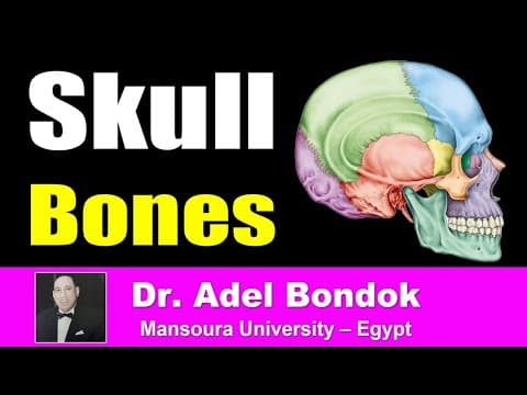
[0:24]I am going to talk about the anatomy of the skull. The skull is viewed from different aspects.
[0:33]Norma Frontalis from the front, this is anterior view of the skull, called norma frontalis.
[0:43]Norma Occipitalis, the view from the back, okay to view the skull from the back. Norma Occipitalis.
[0:53]And then Norma Verticalis viewing the skull from above. And Norma Lateralis viewing the skull from the side, lateral view of the skull.
[1:09]And then Norma Basalis viewing the skull from the base, from below. So Norma Frontalis.
[1:18]Occipitalis, Verticalis, Lateralis, and Basalis. Start by Norma Frontalis.
[1:29]Regarding Norma Frontalis, you should identify the bones, the sutures, the landmarks, and the foramina.
[1:42]Regarding the bones, okay, this is the first bone from above. This is the frontal bone which is the upper part.
[1:52]This is the nasal bone, or nasal bones, one on each side. Okay, this is the zygomatic bone, one on each side.
[2:06]And this is the maxilla forming the upper jaw. And this is the mandible.
[2:13]Regarding the frontal bone, it forms the roof of the orbit, this is the orbital plate. It has maxillary process, and the zygomatic process.
[2:28]Regarding the zygomatic bone, it has frontal process, and maxillary process. Regarding the maxilla, it has frontal process, articulating with the frontal bone.
[2:49]Zygomatic process articulating with the zygomatic bone, and alveolar process forming with the opposite one the alveolar arch. So these are the bones.
[3:02]Regarding the sutures, this is the coronal suture between the frontal and parietal bones.
[3:13]This is the metopic suture. Metopic suture is present in the infant skull between the two halves of the frontal bone.
[3:27]It disappears at the age of 2 to 6 years. The lower part persists in about 8% of adult skulls. The persistence of the metopic suture may be mistaken for a fracture of the frontal bone.
[3:47]Then the frontozygomatic suture, this is the frontozygomatic suture, and this is the frontonasal suture.
[3:59]And this suture between the two nasal bones, the internasal suture. So we identified the bones, we identified the sutures.
[4:11]Now, the bony landmarks and the foramina. The bony landmarks, this is the first one, the frontal eminence.
[4:25]Of course one on each side. And the frontal eminence is the site of center of ossification of the frontal bone.
[4:34]This is the second one, this ridge. This ridge is called the superciliary arch. Superciliary arch above the orbit.
[4:46]Between the two superciliary arches, this elevation, this elevation is called the glabella.
[4:58]And the depression at the frontonasal suture is called the nasion.
[5:06]And then the nasal septum, this is the nasal septum. And these are the nasal conchae, the middle and inferior. Okay, middle nasal concha, and inferior nasal concha.
[5:21]And then this eminence is the canine eminence formed by root of the canine tooth. So we have landmarks, frontal eminence, site of ossification.
[5:35]Superciliary arch, elevation above the orbit, glabella, elevation between the two superciliary arches, nasion, depression at the frontonasal suture.
[5:49]Okay, nasal septum, middle and inferior nasal conchae, and canine eminence. Regarding the foramina, I see four foramina. This is the first one.
[6:03]This is the first one, supraorbital foramen. This supraorbital foramen or notch, it transmits the supraorbital nerve and vessels.
[6:20]This is the second one. Infraorbital foramen. This infraorbital foramen transmits infraorbital nerve and vessels.
[6:34]This is the third one, this foramen, zygomaticofacial foramen. This zygomaticofacial foramen transmits zygomaticofacial nerve and vessels.
[6:49]And this foramen is the mental foramen. This mental foramen transmits mental nerve and vessels. So this is normal frontalis.
[7:01]Let us see this labeled diagram of normal frontalis. Regarding the bones, this is the frontal bone.
[7:12]This is the zygomatic bone. This is the maxilla. Nasal bones, okay. Regarding the landmarks, this eminence is the frontal eminence.
[7:27]This arch, okay, is the superciliary arch. This is the supraorbital foramen. This is the frontozygomatic suture.
[7:40]This is the glabella. And this one is the nasion. This is the coronal suture.
[7:51]This is the zygomatic process of the frontal bone. And this is the frontal process of the zygomatic bone.
[8:04]This is the zygomaticofacial foramen transmitting zygomaticofacial nerve and vessels. Infraorbital foramen transmitting infraorbital nerve and vessels.
[8:19]This is the mental foramen transmitting mental nerve and vessels. This is the canine eminence formed by the root of the canine tooth.
[8:34]My semester 4 students surprised me at the end of an online lecture.
[8:51]Now, Norma Verticalis. This is Norma Verticalis externa from outside. Identify the bones, identify the sutures, and identify the bony landmarks.
[9:07]Regarding the bones, we have three bones here. This is the first one. This is the frontal bone forming the anterior part.
[9:17]This is the occipital bone forming the posterior part, and these two bones are the parietal bones, one on each side. Regarding the sutures, I see three sutures.
[9:33]This is the coronal suture. Coronal suture between the frontal and parietal bones.
[9:41]This is the sagittal suture. Sagittal suture between the two parietal bones. And this is the lambdoid suture between the parietal and occipital bone.
[9:57]Regarding the bony landmarks, the first landmark is this area. This area is called the bregma.
[10:07]So bregma is the meeting of the sagittal and coronal suture. This bregma in the newborn skull, it is the site of anterior fontanelle.
[10:25]This is the anterior fontanelle, diamond-shaped, between the frontal and parietal bones. Okay, so the bregma in the adult skull, anterior fontanelle in the newborn skull.
[10:44]This is the second one here. This is the lambda, and the lambda is the meeting of the sagittal and lambdoid sutures.
[11:00]And the lambda is the site of posterior fontanelle in the newborn skull. And the posterior fontanelle is triangular in shape.
[11:10]The anterior fontanelle ossifies at the age of 18 months, the posterior fontanelle ossifies at the age of 6 months. So regarding the bones of Norma Verticalis externa.
[11:26]Frontal anterior, occipital posterior, parietal bones, one on each side. Regarding the sutures, coronal suture between the frontal and parietal.
[11:37]Sagittal suture between the two parietal bones, and lambdoid suture between the parietal and occipital. Regarding the landmarks, bregma, meeting of the sagittal and coronal sutures.
[11:57]Lambda, meeting of the sagittal and lambdoid sutures. We have this prominence, this is the parietal foramen.
[12:17]Or parietal emissary foramen, it transmits emissary vein between SSS and scalp and this is the parietal eminence which is the site of center of ossification of the parietal bones.
[12:37]This is Norma Verticalis interna. This is an internal view of the vault of the skull. Again, the bones, this is the frontal bone anterior.
[12:50]This is the occipital bone posterior, and these two are the frontal bones, one on each side. Regarding the sutures, this is the coronal suture between the frontal and parietal bones.
[13:04]This is the sagittal suture between the two parietal bones. This is the lambdoid suture between the parietal and occipital bone. Regarding the bony landmarks from inside.
[13:18]Okay, remember, this is the bregma. Yes, bregma. Meeting of sagittal and coronal suture. This is the lambda. Meeting of the sagittal and lambdoid suture.
[13:34]Okay. These grooves, okay, this groove in the midline, this is the groove for superior sagittal sinus, extending from the frontal crest, this is the frontal crest.
[13:48]Okay, so this is the groove for superior sagittal sinus. And these grooves for the middle meningeal vessels. And these depressions, granular foveolae.
[14:04]Granular foveolae are made by the arachnoid granulations. So we have bregma, lambda, groove for superior sagittal sinus.
[14:17]Grooves for the middle meningeal vessels, and granular foveolae made by arachnoid granulations. Okay, this is Norma Verticalis externa.
[14:31]And this is Norma Verticalis interna. The bones, frontal anterior, occipital posterior, parietal on each side. Sutures, coronal suture, sagittal suture.
[14:48]Lambdoid suture. The features, bony landmarks, bregma, lambda, parietal foramen, and parietal eminence site of ossification.
[15:00]Interna, this is the groove for superior sagittal sinus. This is the frontal crest. Coronal suture, sagittal suture, lambdoid suture.
[15:15]And these grooves made by the middle meningeal vessels, and these depressions granular foveolae made by arachnoid granulations.
[15:29]Examination of the skull from the back. This is Norma Occipitalis. Okay, we have to identify the bones, identify the sutures, and the bony landmarks.
[15:43]Regarding the bones, three main bones, on each side, parietal bones, right and left. And this one is the squamous part of the occipital bone.
[15:58]And this one is the mastoid process. Okay. So the bones of Norma Occipitalis, two parietal bones. Squamous part of the occipital bone, and the mastoid process. Regarding the sutures.
[16:17]This is the sagittal suture, and this is the lambdoid suture. Okay, so sagittal suture, lambdoid suture. We have two more sutures. This one, this one.
[16:30]Occipitomastoid, and this one parietomastoid suture. So occipitomastoid suture, and parietomastoid suture. Look at the occipitomastoid suture, there is a foramen here.
[16:45]This is the mastoid foramen. Mastoid foramen. Regarding the bony landmarks. Of course, this is the bregma.
[16:54]Okay, external occipital protuberance. Inion is the prominent point. This is the medial crest external, okay, external occipital crest.
[17:15]This is the bregma. This is the lambda. Lambda meeting of sagittal and lambdoid suture. And this one, this one is called the asterion.
[17:27]And this asterion is the meeting of three bones, parietal, occipital and mastoid process. And this asterion is opposite the sigmoid sinus.
[17:40]And in the newborn skull, it is represented by the posterolateral fontanelle. It ossifies at the age of 3 months. In the clinical importance or neurosurgical importance, it overlies the sigmoid sinus.
[17:58]Asterion. I will go back to the asterion later on.
[18:05]Three nuchal lines. This is the superior nuchal line. This is the inferior nuchal line, and there is a small one here, called the highest nuchal line. So the three nuchal lines, highest, superior, and inferior.
[18:24]This is the mastoid notch or digastric notch because it gives attachment to the posterior belly of the digastric muscle.
[18:37]This is the mastoid foramen. This mastoid foramen transmits artery and vein. The vein is emissary vein between the sigmoid sinus and occipital veins. And the artery is the meningeal branch of the occipital artery.
[18:57]So what are the structures passing through the mastoid foramen? Artery and vein. The artery is the meningeal branch of the occipital artery, and the vein is emissary vein between the sigmoid sinus and occipital veins. So this is Norma Occipitalis.
[19:19]Okay, again, this is Norma Occipitalis. Okay, let us identify the bones. Two parietal bones. Squamous part of the occipital bone.
[19:32]And this is the mastoid process. Okay. This is the lambda. Okay. Meeting of sagittal and lambdoid suture.
[20:41]This is the asterion. Asterion is meeting of three bones. Parietal, occipital and mastoid process. And this asterion is opposite the sigmoid sinus.
[20:56]And in the newborn skull, it is represented by the posterolateral fontanelle. It ossifies at the age of 3 months. This is the mastoid foramen. Okay. So this is the sagittal suture. This is the lambdoid suture. Just reminding you, the mastoid foramen transmits artery and vein.
[20:25]The artery is the meningeal branch of the occipital artery. The vein is emissary vein between the sigmoid sinus and occipital veins.
[31:03]My Semester 4 students surprised me at the end of an online lecture.
[31:25]Now, what are the four fontanelles of the newborn skull? Four fontanelles. This is the first one. This is the anterior fontanelle.
[31:37]Anterior fontanelle between frontal and parietal bones in the adult skull. It's represented by the bregma. It ossifies at the age of 18 months. This is the posterior fontanelle.
[31:58]Posterior fontanelle between the parietal and occipital bones represented by the lambda. And it ossifies at the age of 6 months. This is the anterolateral fontanelle.
[32:14]Anterolateral fontanelle between four bones, frontal, parietal, GWS and temporal bones represented by the pterion. And it ossifies at the age of 3 months. And this is the asterion.
[32:30]Posterolateral fontanelle, the posterolateral fontanelle between three bones, parietal, occipital and temporal. It is represented by the asterion in the adult skull, and it ossifies at the age of 3 months. So these are the four fontanelles, anterior, posterior, anterolateral, posterolateral in the adult skull.
[32:53]Bregma, lambda, pterion, and asterion. Regarding Norma Basalis or the base of the skull, it will be discussed in a coming video. So wait for that video. And thank you very much.
[33:12]Best wishes, and good luck. And see you very soon with a video on the base of the skull.


